


A Guide to Clinical Differential Diagnosis of Oral Mucosal Lesions
COURSE NUMBER: 110
Credit Hours:
4 Hour(s)
SHARE
The primary goal of this course is to help dental professionals learn the process of clinical differential diagnosis of diseases and lesions of the oral mucosa. This course also includes both an interactive and downloadable decision tree to...
(Use this feature to create assignments for your students and staff.)
Overview
The primary goal of this course is to help dental professionals learn the process of clinical differential diagnosis of diseases and lesions of the oral mucosa. This course also includes both an interactive and downloadable decision tree to assist in the diagnosis.
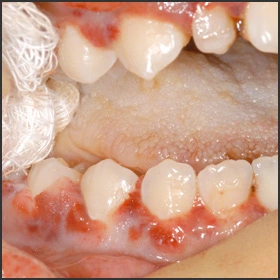
Oral pathology is a visual specialty, and clinical images can facilitate your learning the clinical features of oral mucosal lesions. Several atlases are recommended in the Additional Resources section of this course. The textual material in this course is designed to be used with “The Oral Pathology Image Database (Atlas).”
Please note that lesions or diseases discussed in the textual material that have clinical images available on “The Oral Pathology Image Database” are designated with *.
Intended Audience:
Dental Assistants, Dental Hygiene Students, Dental Hygienists, Dental Students, Dentists, Dental Assisting Students
Date Course Online:
Jun 3, 2002
Last Revision Date:
Mar 25, 2020
Course Expiration Date:
Mar 24, 2023
Cost:
Free
Method:
Self-instructional
AGD Subject Code(s):
Learning Objectives
Upon completion of this course, the dental professional should be able to:
Classify oral lesions into surface lesions and soft tissue enlargements using a decision tree (flowchart).
Describe the clinical features that are characteristic of each class of oral mucosal lesions in the decision tree, including:
White surface lesions - epithelial thickening, surface debris, and subepithelial change
Generalized pigmented surface lesions
Localized pigmented surface lesions - intravascular blood, extravascular blood, melanin pigment, and tattoo
Vesicular-ulcerated-erythematous surface lesions - hereditary, autoimmune, viral, mycotic, and idiopathic
Reactive soft tissue enlargements of oral mucosa
Benign tumours of oral mucosa - epithelial, mesenchymal, and salivary gland
Malignant neoplasms of oral mucosa
Cysts of oral mucosa
Describe the characteristic or unique clinical features of the most common and/or important diseases of the oral mucosa.
Perform a step-by-step clinical differential diagnosis, using the decision tree, for patients with oral mucosal lesions.
Disclaimers
Participants must always be aware of the hazards of using limited knowledge in integrating new techniques or procedures into their practice. Only sound evidence-based dentistry should be used in patient therapy. Note: Registration is required to take test.
Submission Information
Recognition

Approved PACE Program Provider
THE PROCTER & GAMBLE COMPANY
Nationally Approved PACE Program Provider for FAGD/MAGD credit.
Approval does not imply acceptance by any regulatory authority or AGD endorsement.
8/1/2021 to 7/31/2027
Provider ID# 211886
(Use this feature to create assignments for your students and staff.)