


Digital Imaging in Dentistry: Intraoral, Extraoral, and 3D Technology
Course Number: 512
Course Contents
Characteristics of Digital Imaging Detectors
Contrast resolution is an important characteristic in analog and digital imaging. The ability to differentiate amongst densities is vital for the dentist when diagnosing diseases and conditions. Current digital detectors capture data from 8-16 bits. The bit depth is at a power of 2. This means that the detectors can capture 256 (2.8) or higher of gray levels. A high contrast-to-noise ratio provides the closely spaced gray values needed for higher contrast when diagnosing diseases and conditions. Current computer monitors can detect 242 levels of gray, and the human eye can detect about 60 gray levels (Figure 25). The level of ambient light in the treatment rooms do not affect digital image viewing compared to conventional radiographs on a view box.5,9 One recent research study found a degradation in spatial resolution after the same PSP plates had been used 48 times. This finding is important, as PSP plates have been shown to have low spatial resolution compared to other intraoral imaging receptors.2
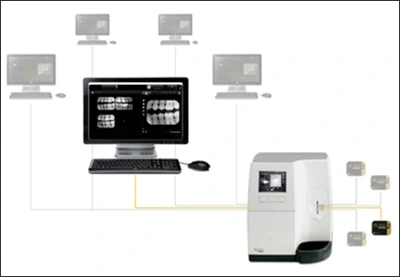
Figure 25. Digital Imaging Hardware and Software.
Image Source: Carestream Dental, LLC, Atlanta, GA.
Spatial resolution provides the fine detail needed to view an image. Resolution can be measured in units of line pairs per mm. At least two columns of pixels are required for the line pairs to create light and dark spaces. Some of the newer solid-state sensors have excellent resolution that is helpful in diagnosing incipient lesions. Although the sensors are thicker than other manufacturers, the contrast is exceptional. The loss of spatial resolution with PSP plates is determined by the construction of the plate and the physical design and laser scanner settings.5,9 Signs of malfunction of the laser scanner include lines across the image due to scanner belt slippage or photomuliplier tube malfunction, inconsistent shading and enlarged window display if the size 2 image due to photomultiplier tube filter malfunction. Several research studies have indicated replacing PSP plates after 50-60 uses. Some of the primary reasons to replace PSP plates are the presence of scratches on the plates and loss of spatial resolution. The use of aluminum step wedges and contrast wells in research studies was helpful in determining contrast changes.
2Image enhancement is used to view digital images. For example, digital systems can magnify images four times the original size to diagnose dental diseases and conditions. Many digital systems software includes a measurement tool for endodontic or dental implant therapies. Some of the digital systems provide split screen technology allowing the dental provider the ability to compare multiple images together and color for contrast during diagnoses.