


Digital Imaging in Dentistry: Intraoral, Extraoral, and 3D Technology
Course Number: 512
Course Contents
Infection Control with Digital Imaging
As with any type of radiology imaging, infection control methods should be utilized to not cross contaminate when exposing images. With digital imaging, barriers must be placed on the sensors. Sensors cannot be sterilized. There are many types of plastic barriers available for indirect phosphor plate and hard sensors used in direct digital imaging. The wired direct sensors will need barriers over the fiber optic cables and hard sensor. The direct wireless sensor will also need a barrier (Figures 19-22). Many of the digital image receptors are autoclavable. There are also some digital receptors that are autoclavable, but have disposable intraoral components (Figure 23). It’s recommended you talk to your dental sales representative and your colleagues as to what they recommend with your type of digital imaging system, as well as finding samples at regional and national dental conferences to try out. When working with phosphor plates, each sensor must be wrapped in a plastic barrier envelope to protect the sensor from saliva (Figure 24). If the sensor becomes contaminated, check manufacturer instructions to see how the sensors can be disinfected. If contaminated, the CDC recommends an EPA-registered intermediate-level disinfectant after removing the barrier and before using the sensor on the next patient.
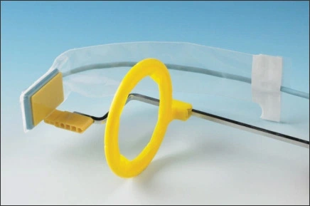
Figure 19. Autoclavable Digital Receptor with Barrier Covering Sensor and Cable.
Image Source: Schick by Sirona, Sirona Dental, Inc., Long Island City, NY.
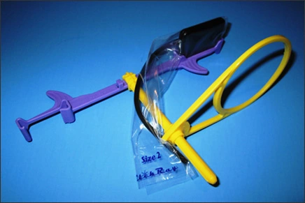
Figure 20. Autoclavable Digital Receptor with Barrier.
Image Source: Clik Ray, Clik Tech, LLC, Scottsdale, AZ.
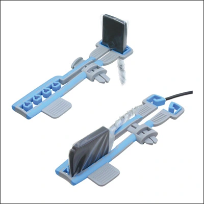
Figure 21. Autoclavable Digital Receptor with Sensor Barriers.
Image Source: Dentsply International, York, PA.
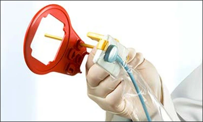
Figure 22. Autoclavable Digital Receptor with Sensor Barriers.
Image Source: Schick by Sirona, Sirona Dental, Inc., Long Island City, NY.
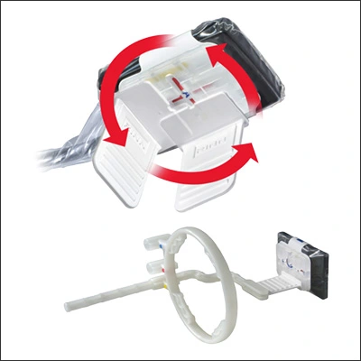
Figure 23. Autoclavable Digital Receptor with Disposable Intraoral Component.
Image Source: Dentsply International, York, PA.
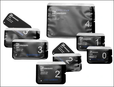
Figure 24. Indirect Photo-stimulable Phosphor Plates with Disposable Barrier Envelopes.
Image Source: Henry Schein Air Techniques, Inc., Melville, NY.