


Cone-Beam Computed Tomography (CBCT) Applications in Dentistry
Course Number: 531
Course Contents
Orthodontics
CBCT has been used in orthodontics due to the capacity of imaging a large field of view that includes all the landmarks for cephalometric analysis. For analysis of skeletal jaw relationships, the three-dimensional image needs to include the skull base, facial bones, and facial soft tissues. By exporting the DICOM files into specialized software programs, orthodontists are able to perform cephalometric analysis and treatment planning. A CBCT study will be helpful in an orthodontic case with proposed orthognathic surgery.17
Another application of CBCT in the field of orthodontics is to find the exact location of any impacted teeth, along with the relationship of adjacent anatomical structures. This information can assist the clinician in determining the best approach for each case. If root resorption of teeth is present due to impacted teeth, the extent of the resorption may be evaluated through CBCT imaging (Figure 14). The main advantage of CBCT over other dental imaging is the cross-sectional view without superimposition, where the buccolingual position of the roots can be assessed. Despite the benefits and advantages of CBCT studies, it should not be routinely used for all orthodontic patients.17,18
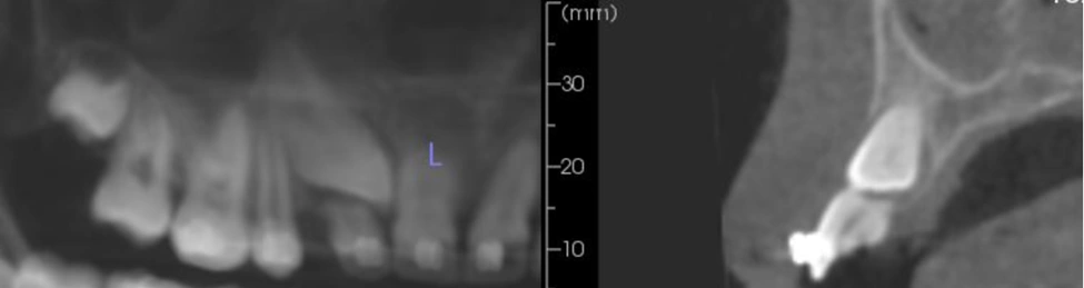
Figure 14. Impacted tooth #6 with apical root resorption on tooth #7