


Panoramic Radiographs: Technique & Anatomy Review
Course Number: 533
Course Contents
Patient Positioning
In order to obtain diagnostically useful images, patients must be positioned carefully within the image layer or focal trough, which is a three-dimensional curved zone (Figure 5). Structures found within the image layer will be reasonably well-defined.5 The patient must be positioned correctly so that the proper structures are aligned within the image layer.
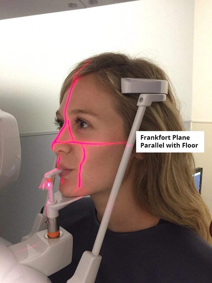
Figure 5.
Example of correct patient positioning with the tongue pressed against the palate, teeth in the groove of the bite-block, and the indicator light for the midsagittal plane centered and perpendicular to the floor.
If patient positioning is incorrect, errors are likely to occur. Patient positioning errors are the most common type of error when performing panoramic radiography.8 For instance, in a study evaluating 460 panoramic radiographs, careless head positioning accounted for 38% of the errors.5 Patient positioning errors accounted for 85% in a sample of 1,813 panoramic radiographs.5
The most common patient positioning error occurs when the tongue is not placed close enough to the palate.5 This may be due to the patient misunderstanding the instructions and only placing the tip of their tongue on the palate. Incorrect positioning of the tongue creates radiolucency near the apices on the maxilla, which makes diagnosis of periodontitis and root resorption challenging.5
It is helpful to note that each manufacturer provides specific operation instructions in the manual that accompanies the unit. It is worthwhile for each team member to become acquainted with the contents of the manual. While the instructions make panoramic imaging easy to perform well, it is equally as easy to perform badly when manufacturers’ instructions are not followed.2 Proper patient positioning (Table 4) will help reduce the possibility of errors in panoramic imaging.
| Standing/Sitting |
|
| Mouth position |
|
| Midsagittal Plane |
|
| Frankfort Plane |
|
| Tongue |
|
| Lips |
|
| Eye |
|