Panoramic Radiographs: Technique & Anatomy Review
Course Number: 533
Course Contents
Review of Normal Anatomical Landmarks and Variations
It is important to understand the landmarks normally seen on panoramic images in order to prevent misdiagnosis of a radiopaque or radiolucent area. For the purposes of this course, we will focus on the structures that are most commonly viewed in panoramic images. For additional information, a review of the anatomic structures can be found in the article by Farman2 and the text by Iannucci & Howerton.1
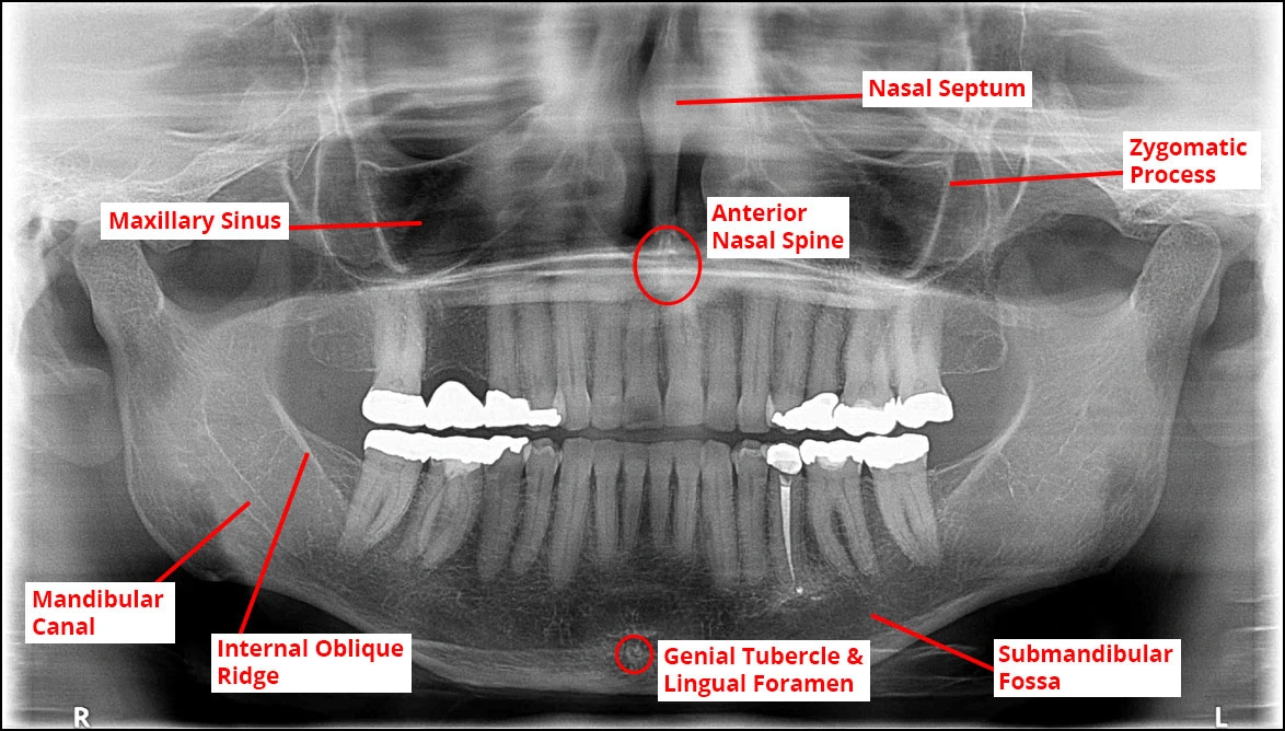
Figure 2 below, includes many of the normal anatomical landmarks that will be visible on a diagnostic panoramic image. The maxillary sinuses are radiolucent and can be found bilaterally on either side of the nasal septum. The zygomatic process is a vertical, radiopaque line that forms the anterior portion of the zygomatic arch (cheekbone). In the mandibular region, the mandibular canal is bordered by two radiopaque lines and it houses the inferior alveolar nerve. The internal oblique ridge is a bony landmark that may be palpated during an inferior alveolar nerve block. At the midline of the mandible, there is a radiolucent lingual foramen that is bordered by genial tubercles, which are radiopaque. Finally, the submandibular fossa is a radiolucent depression that houses the sublingual gland.
Figure 2. Normal Anatomical Landmarks.3
(Refer to the glossary for the definition of each structure shown).
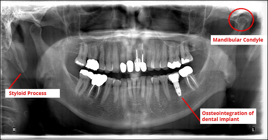
In Figure 3 below, the patient’s chief complaint was popping near the temporomandibular joint (TMJ). The panoramic image indicated a flattened condyle and significant wear of the glenoid fossa of the temporal bone due to constant force from bruxism and clenching. It was also noted that the patient had very pronounced styloid processes (bilaterally).
Figure 3. Example of Pathology and Variations of Normal.
(Refer to the glossary for the definition of each structure shown.)
Image source: Courtesy of AB & Dr. Iwata


