Digital Imaging in Dentistry: Intraoral, Extraoral, and 3D Technology
Course Number: 512
Course Contents
Intraoral Digital Imaging Procedures
Step-by-step digital imaging procedures vary by manufacturer. As with all aspects of dentistry, quality assurance is also vital with digital imaging. The National Council on Radiation Protection and Measurements Report 145 indicates there must be a written protocol for dental offices for their image receptor systems.7 One research study found that many dental offices were using incorrect exposure settings resulting in lower diagnostic capabilities and higher radiation dosage. 8 Despite proper care and handling of PSP plates, loss in spatial resolution may occur. To reduce PSP plate deterioration resulting in surface damage and contamination, PSP plates should be handled carefully and cleaned according to manufacturer instructions.2
It is strongly recommended that the dental practice schedule more than one in-office training with clinical dental providers on the computer hardware and software, and the digital imaging equipment. The dental practice might need more time with the trainer than what is part of the digital image package pricing. The key to any practice is efficiency of time and energy for both the dentist and their staff. The more that clinical staff can troubleshoot digital imaging techniques, the more time they can dedicate to other dental practice treatment.
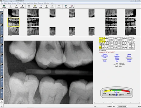
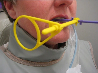
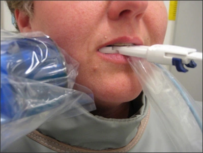
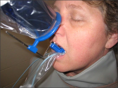
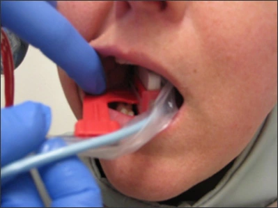
The sequencing of how the clinical provider exposes bite-wing and periapical digital imaging can be the same as traditional films. However, the dental manufacturer representative who sets up your digital imaging system will discuss the number of digital images you wish to use for full-mouth series and bite-wing series, as well as sequencing to see what you prefer (Figure 14). These in-office trainings, without radiation, allow staff to troubleshoot placement with different receptors in each other’s mouths. Figures 15-18 show the clinical provider using different autoclavable direct sensor receptors. Note the plastic barrier over the fiber optic cable.
Figure 14. Digital Full-mouth Series.
Image Source: Dentsply International, York, PA.
Figure 15. Bite-wing Direct Digital Imaging.
Image Source: Prof. Willie Leeuw, MS; Brandy Spaulding, MS; Sarah Pulver, MS; Indiana University, Fort Wayne, IN.
Figure 16. Posterior Periapical Direct Digital Imaging.
Image Source: Prof. Willie Leeuw, MS; Brandy Spaulding, MS; Sarah Pulver, MS; Indiana University, Fort Wayne, IN.
Figure 17. Anterior Periapical Direct Digital Imaging.
Image Source: Prof. Willie Leeuw, MS; Brandy Spaulding, MS; Sarah Pulver, MS; Indiana University, Fort Wayne, IN.
Figure 18. Bite-wing Direct Digital Imaging.
Image Source: Prof. Willie Leeuw, MS; Brandy Spaulding, MS; Sarah Pulver, MS; Indiana University, Fort Wayne, IN.


